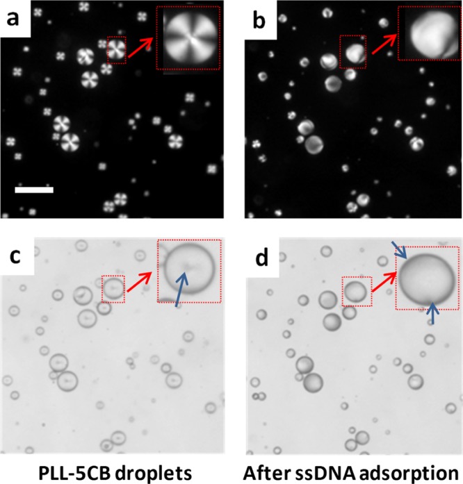
Figure 1.

Polarized optical and corresponding bright-field micrograph images of PLL-coated LC droplets in contact with: (a,c) 0 μM ssDNA and (b,d) 30 μM ssDNA. The LC droplets were in radial states (a,c) before but transitioned to a bipolar/preradial state (b,d) 10 s after the addition of ssDNA. The insets within (a–d) indicate the higher magnification version of the red arrow-marked LC droplet. Blue arrows in (c,d) indicate the point defect in the center of a radial droplet and two defects at the poles of a bipolar droplet, respectively. Scale bar = 50 μm.
