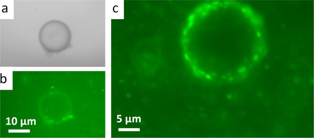
Figure 8.
Bright-field (a) and corresponding epi-fluorescence microscopic image (b) of a PLL–5CB droplet after adsorption of 30 μM FAM-tagged fluorescent ssDNA in the presence of excess PLL. Epi-fluorescence microscopic image (c) corresponds to another PLL–5CB droplet after adsorption of 30 μM FAM fluorescent ssDNA clearly showing the presence of fluorescent polyplexes at the surface of the droplet.

