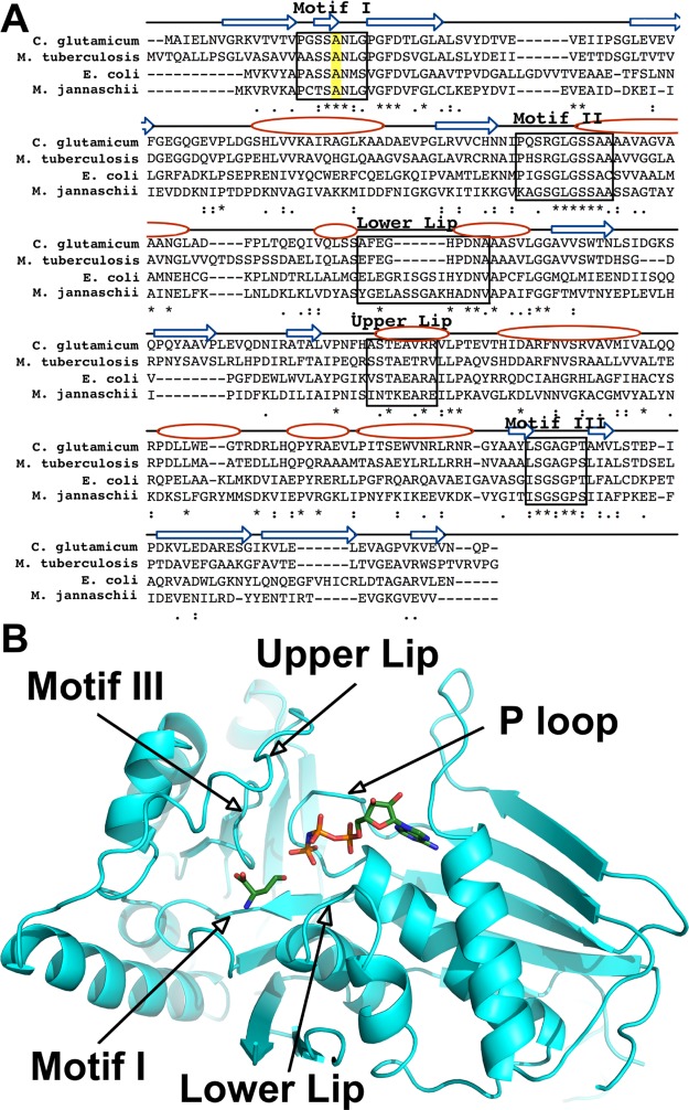
Figure 2.
(A) Alignment of C. glutamicum homoserine kinase (CglThrB) sequence with those of E. coli, M. jannaschii, and M. tuberculosis using CLUSTAL O (1.2.4). Conserved residues are shown with asterisk (*), residues with strongly similar properties are shown with a colon (:), and residues with weakly similar properties are shown with a period (.). The conserved Ala residues in motif I are highlighted in yellow. The arrows represent β-strands, whereas the ovals represent α-helices. (B) Structure of C. glutamicum ThrB (CglThrB-WT) with modeled l-homoserine and AMP-PNP based on the structure of M. jannaschii ThrB, with these compounds bound in the active site.

