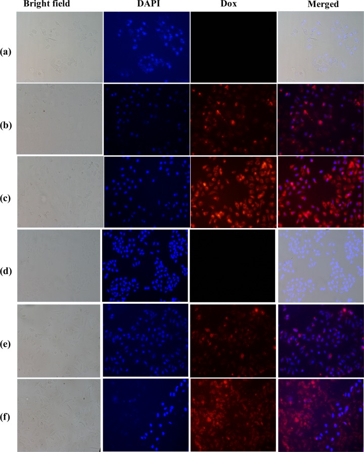
Figure 8.
Localization of Dox by MCM-allylCalix-Dox using fluorescence microscopy: (a)–(c) for MCF7 and (d)–(f) for HeLa cells. (a, d) Corresponding cells alone as controls. (b, e) Cells treated with 50 μg/mL MCM-allylCalix-Dox. (c, f) Cells treated with 100 μg/mL MCM-allylCalix-Dox. Red fluorescence indicates Dox, and blue indicates 4′,6-diamidino-2-phenylindole (DAPI) stain. The columns from left to right correspond to bright field, DAPI, Dox, and merged.

