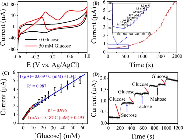
Figure 6.
(A) CVs of Pt/HCS in N2-saturated 0.1 M PBS (pH = 7.4) solution with and without 50 mM glucose at a scan rate of 5 mV s–1. (B) Amperometric response of Pt/HCS-modified GCE to successive addition of glucose at the potential of 0.6 V in N2-saturated 0.1 M PBS solution (pH = 7.4); the inset shows the amplified current signal at low concentrations of glucose. (C) The corresponding calibration curve of Pt/HCS for the detection of glucose. (D) The current response of Pt/HCS to the addition of 2 mM glucose and 2 mM different interfering analytes (sucrose, lactose, and maltose) in 0.1 M PBS solution (pH = 7.4) at 0.6 V.

