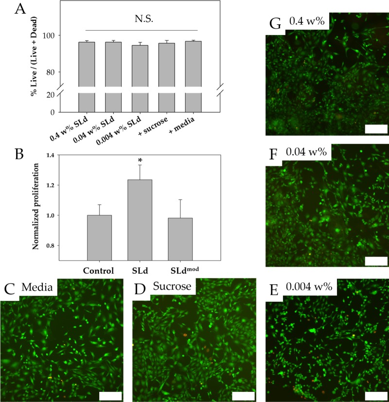
Figure 4.
Cytocompatibility and DPSC proliferation. (A) Viability of fibroblasts cultured with SLd at different concentrations (0.004–0.4 wt % w/v), media with formulation solution (298 mM sucrose), or media-only are shown along with their representative live/dead images (C–G). (B) Dental pulp stem cells demonstrated stronger proliferation (CCK-8) compared to no treatment of 0.004 wt % SLd or treatment with a modified variant (SLdmod) with similar charge density (n = 8, *p < 0.01). Scale bar (C–G): 200 μm.

