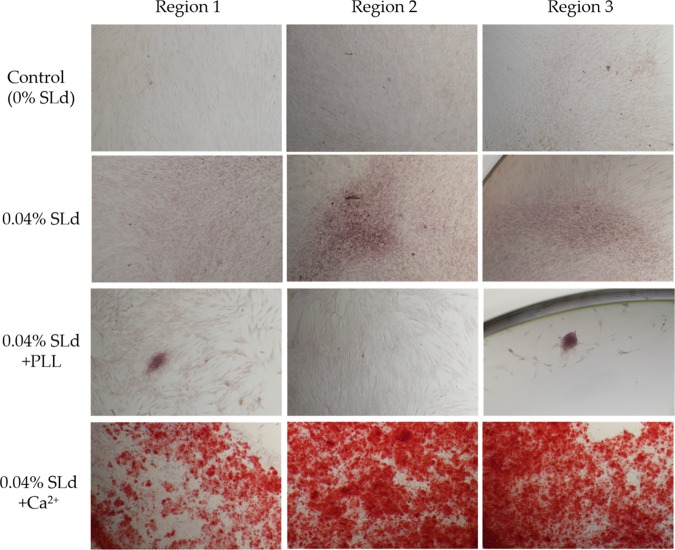
Figure 5.
Calcium phosphate deposition in response to SLd treatment of DPSCs (representative regions from independent cultures). SLd hydrogel shows appreciable red staining either alone or in combination with poly-l-lysine (PLL) compared to media-only controls. The addition of Ca2+ to the SLd formulation results in significantly more observable calcium phosphate deposition, as observed with Alizarin red staining.

