
Fig. 8.
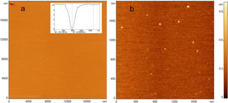
(a) AFM image of a 20×20 μm area of POPS SLB prepared by incubating 0.5 mg/mL lipid in phosphate buffer at 60°C for 1 h. A single packing defect is visible in the upper left-hand corner of the scanned image. Cross-section profile in the inset demonstrates the expected height for a packing defect. (b) α-Syn aggregates on POPS bilayer after 1 h