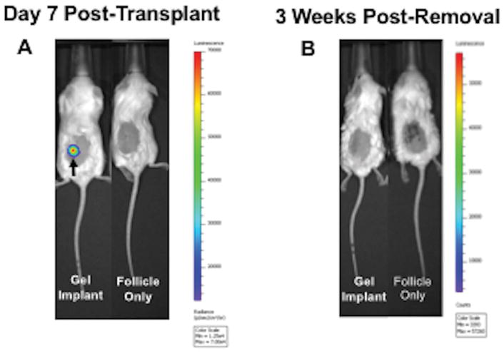
Figure 6. In Vivo Imaging of NSG Mice Pre- and Post-Removal of Cancer-Laden Alginate Hydrogels.
(A) Representative image of NSG mouse with subcutaneously-transplanted alginate hydrogel containing 200 MDA-MB-231 cells expressing luciferase (imaged Day 7 post-transplant). The presence of cancer cells in the hydrogels was confirmed by the positive luminescent signal (signal detection is 600–60,000 counts). The hydrogel implant at Day 7 is denoted with a black arrow. For the control group, mice transplanted with only ovarian follicles were imaged. Cancer cells were present only in the gel implant. (B) Mice were imaged 3 weeks after hydrogels removal to assess cancer cell presence. Cancer cells were not detected in experimental mice that had cancer-laden hydrogels removed at Day 7. Cancer cells were not detected in negative controls as well. 360 ovarian follicles were also incorporated into transplanted alginate hydrogels. n=4 per group.

