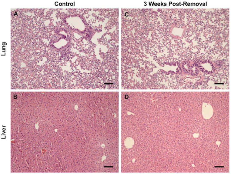
Figure 7. Absence of Metastatic Lesions in Liver and Lungs of Recipient Mice 3 Weeks Post-Removal of Hydrogel.
Representative image of (A) liver from naïve NSG control (n=3), (B) lung from naïve NSG control (n=3), (C) liver from recipient mouse with 200 cancer cells in hydrogel (n=4), and (D) lung from recipient mouse with 200 cancer cells in hydrogel (n=4). Lung and liver tissue removed at 3 weeks did not contain any cellular abnormalities or metastatic growths according to staining with hematoxylin and eosin (H&E). Scale bar: 100 μm.

