
Figure 2.
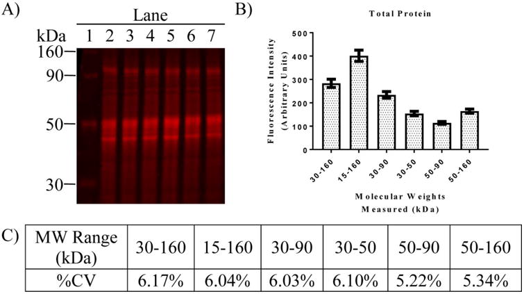
Comparative analysis of the variability in TP signal across different ranges of MW for samples of regional brain homogenates. (A) Image of PVDF membrane showing TP stain of six different hippocampal homogenates for MWs ranging from 30-160 KD. (B) Analysis of fluorescence intensity for different ranges of molecular weight from 12 samples run on a single gel (not shown). Bars represent the mean fluorescence intensity ± SEM for each MW range. (C) %CV calculated from the same 12 samples summarized in B. Note the consistent and low variability in TP measurements across multiple MW ranges.