Figure 3.

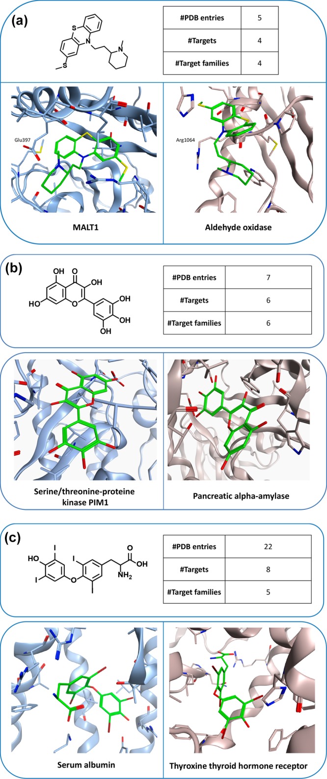
Multifamily ligands and X-ray structures. In (a–c), exemplary ligands and X-ray structures of their complexes with targets from different families are shown. For each ligand, the total number of complex X-ray structures, the number of PDB targets, and the number of families from which these targets originated are reported. In the X-ray structures, bound ligands are shown in stick representation with standard atom coloring.
