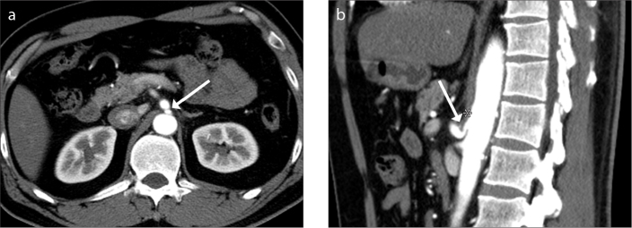
Figure 1 a, b.
Representative case of a 48-year-old man diagnosed with median arcuate ligament syndrome (MALS). Contrast-enhanced arterial phase axial CT image (a) shows abrupt slit-like luminal narrowing (arrow) in the proximal celiac trunk at the fibrous attachment site of the diaphragmatic crura. Sagittal reconstructed arterial phase CT image (b) reveals hooked appearance of the proximal celiac trunk (arrow) due to MAL (asterisk) indenting the superior border of the celiac trunk.

