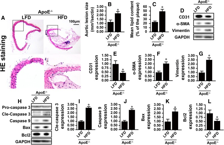
Fig. 1.
The atherosclerosis is developed in HFD mice. In the current study, ApoE−/− mice were treated with low-fat diet (LFD) and/or high-fat diet (HFD). a, b After 12 weeks, H&E staining was performed on aortic sinus and the results demonstrated a significant increase in aortic lesion size, shown by representative images (a) and by quantification (b). c The quantification of lipid content by Oil-red-O-staining on aortic sinus. d–g The proteins in aortic arch were isolated and analyzed the expression of CD31, α-SMA, and vimentin via western blotting. h–l The changes of pro-apoptotic proteins in aortic arch. *P < 0.05. n = 10/group

