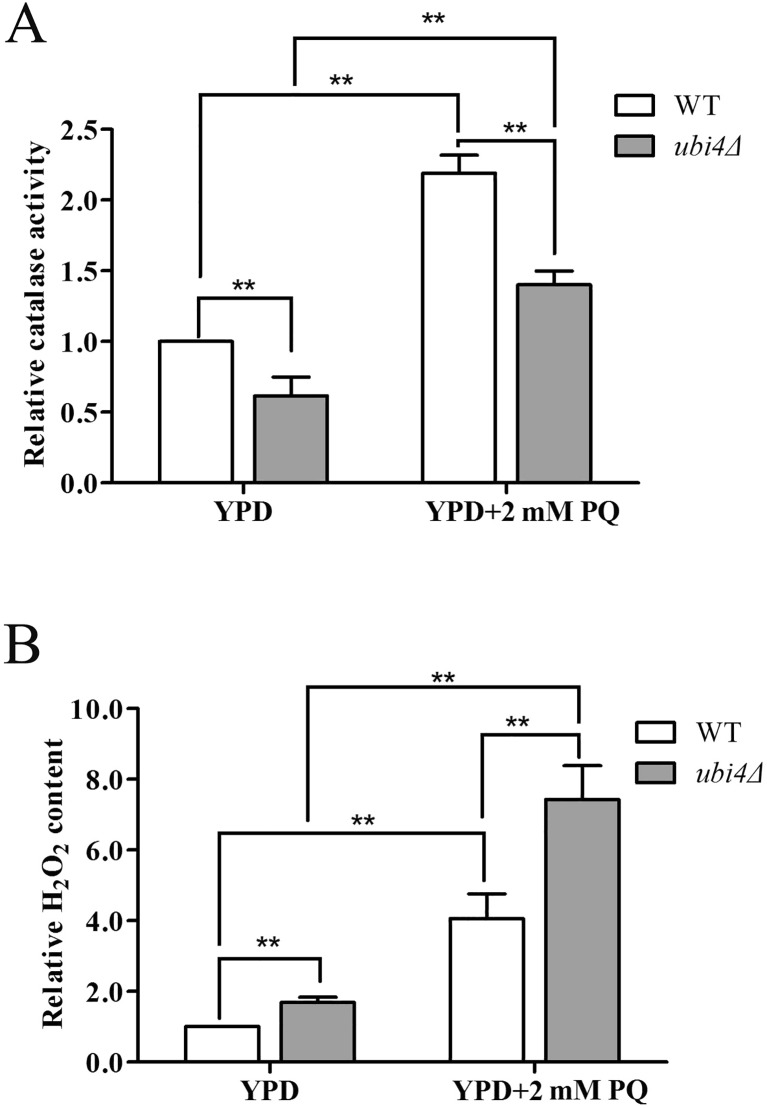
Fig. 3.
Ubi4Δ cells have decreased catalase activity. Catalase activity assay of the wild-type and ubi4Δ cells (a) under the unstressed and PQ-stressed conditions. Intracellular H2O2 levels of the wild-type and ubi4Δ cells (b) under the unstressed and PQ-stressed conditions. *p < 0.05 was considered to indicate a significant difference, and **p < 0.01 was considered to indicate a highly significant difference

