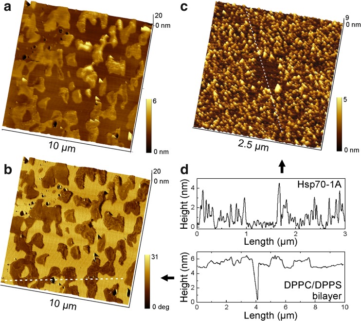
Fig. 1.
a AFM topography image of a phase separated DPPC/DPPS (80:20) bilayer on mica, captured in intermittent contact mode in buffer solution at ambient temperature. The DPPS phase emerged as islands with a corrugated surface from the surrounding smooth DPPC phase. b The overlay of the topography with the phase shift image shows DPPS domains as dark contrast. c Human Hsp70-1A adsorbed on ultra-flat mica. In the center of the image, proteins were removed by AFM lithography (square). d Cross sections along the indicated lines in b and c illustrate typical height profiles

