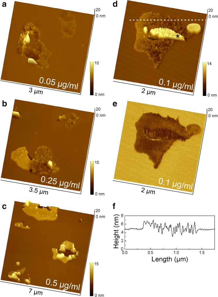
Fig. 2.
DPPC/DPPS (80:20) bilayer after incubation with Hsp70-1A for 30 min. Molecules selectively associated with DPPS domains of the bilayer and formed clusters in a concentration-dependent manner. a Protein concentration of 0.05 μg/ml, b 0.25 μg/ml, and c 0.5 μg/ml. At a maximum concentration of 0.5 μg/ml, Hsp70-1A was densely packed and large membrane defects appeared in the center of each protein cluster. d Topography and corresponding phase shift image (e) at a protein concentration of 0.1 μg/ml clearly show that the protein associated only with DPPS domains. Note that part of the protein layer (marked with an asterisk *) is covered by an additional bilayer patch. f Representative height profile across a section marked in subfigure (d) for approximation of the height of inserted proteins

