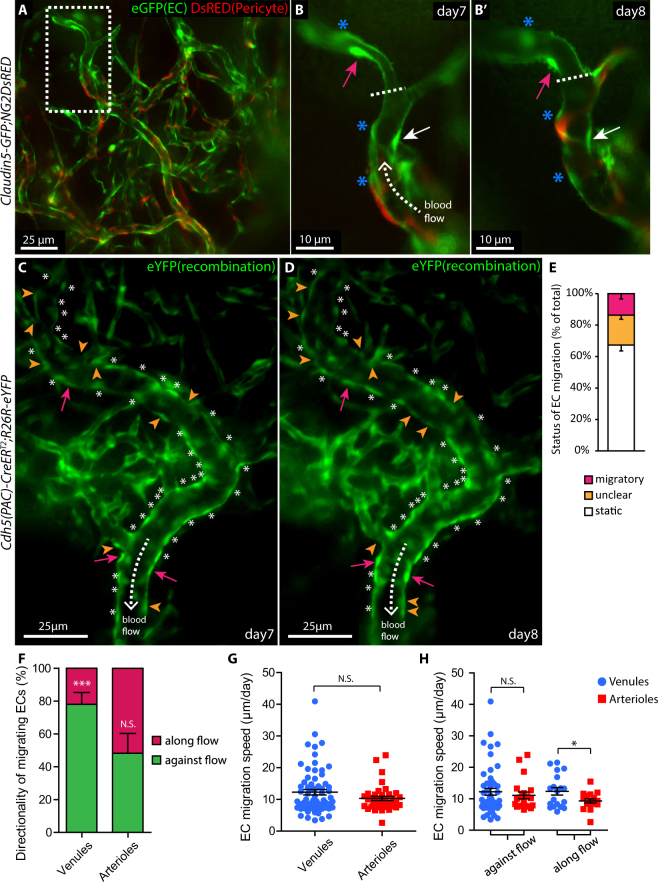
Figure 2.
Time-lapse intra vital microscopy reveals specific EC migratory behaviour in correlation with blood flow direction and arterio-venous identity. (A) Live imaging of the remodelling vasculature of Claudin5-GFP:NG2DsRED mice at day 7 after suture implantation allows for simultaneous recording of ECs (green) and pericytes (red). The dashed box area is magnified in (B). (B,B’) Vessels imaged day 7 and 8 respectively, expose migrating (magenta and white arrows) and static ECs (asterisks), in relation to flow direction (dashed arrow). The vessel bifurcation (dashed line) serves as reference point. (C,D) Venules imaged day 7 and 8, following suture implantation, reveal migratory ECs (magenta arrows), static ECs (asterisks), ECs with non-identifiable direction (unclear, orange arrowheads) as well as blood flow direction (dashed arrows). (E) Percentage of total ECs in a migratory, static or unclear state, within lumenized vessels (n = 6 mice). (F) Fraction of ECs, within the group defined as migratory, that migrate along or against flow direction, in lumenized venules or arterioles. ***p < 0.001. (G) EC migration speed (all directions, pooled data from 9 mice) within lumenized venules (n = 69) and arterioles (n = 38). (H) EC migration speed within lumenized venules and arterioles in relation to flow direction. *p < 0.05.

