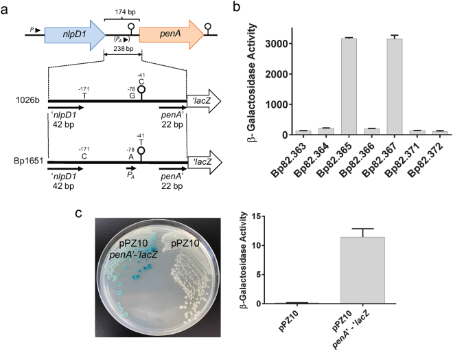
Figure 4.
Promoter activity in native and genetically engineered nlpD1-penA intergenic regions. (a) Design of reporter lacZ reporter gene constructs. The indicated 238 bp DNA fragments shown by the thick lines (plus flanking SpeI and HindIII restriction sites) were obtained from PCR fragments amplified from 1026b and Bp1651 genomic DNA. The indicated −41C, −78G and −171T nucleotides present on the 1026b fragment were individually changed to −41T, −78A and −171C present in Bp1651. Lastly, a 532 bp DNA fragment (plus flanking SpeI and HindIII restriction sites) containing a 336 bp instead of a 42 bp nlpD1 terminus was obtained from a PCR fragment amplified from 1026b genomic DNA. These fragments were cloned into a mini-Tn7-lacZ transcriptional fusion vector and integrated into the Bp82.27 chromosome. (b) β-Gal activity in strains harboring the single-copy lacZ reporter constructs. The strains resulting from chromosomal integration were Bp82.363 (empty vector), Bp82.365 (1026b fragment), Bp82.365 (Bp1651 fragment), Bp82.366 (1026b fragment with −41T), Bp82.367 (1026b fragment with −78A), Bp82.371 (1026b fragment with −171C) and Bp82.372 (1026b fragment with a 331 bp instead of 42 bp nlpD1 3′ terminus. β-Gal activities were measured in triplicate on three separate days and are expressed in Miller units. Error bars indicate standard deviation from the mean. (c) The PA promoter is active in E. coli. A plasmid was constructed that contains a penA’-‘lacZ translational fusion under transcriptional and translation control of the 174 bp 1026b IR with the -78 G to A transition. The resulting pPZ10 penA’-‘lacZ thus contains the PA promoter. The first 7 amino acids of PenA are fused in-frame to LacZ. The resulting pPZ10 penA’-‘lacZ and the empty pPZ10 vector were transformed into strain DH5α. β-Gal activity was assessed by plating on an LB plate with X-Gal indicator (left) or by measuring hydrolysis of ortho-nitrophenyl-β-D-galactopyranoside (ONPG) in culture grown cells (right). The ONPG β-Gal assays were performed in triplicate and activity is expressed in Miller units. Error bars indicate standard deviation from the mean. The plate picture is a minimally processed section of an image of an agar plate shown in Fig. S6.

