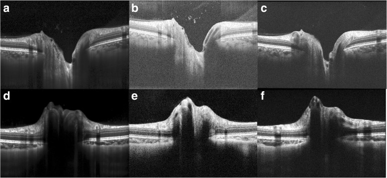
Fig. 3.
Approximately 6-mm OCT scans centered over the optic nerve (taken from radial scan patterns). (a, d) show radial scans taken on the Heidelberg Spectralis OCT device. (b, e) show radial scans acquired on the Optovue Avanti OCT device. (c, f) show radial scans taken on the Zeiss Cirrus OCT device. (a-c) are images of a non-swollen optic nerve while (d-f) are images of a swollen optic nerve in a subject with papilledema due to elevated intracranial pressure

