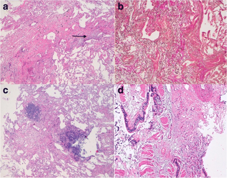
Fig. 3.

(Top: PPFE left: a right: b lower: airway-centered FE left: c right: d). a Markedly thickened visceral pleura and prominent sub pleural fibrosis comprised of a homogenous mixture of elastic tissue and dense collagen. Some fibroblastic foci are evident at the edge between the fibrotic area and normal lung parenchyma (H&E, low power). b The elastic tissue is clearly marked by a specific stain (Verhoeff-van Gieson stain, mid power). c. Fibroelastotic tissue sited in the peribronchiolar acinar area with constructive bronchiolitis (only pulmonary artery branched are identifiable) and focal nodular lymphocytic inflammation. The rest of the lung parenchyma is spared. (H&E, low power). d Another sample showing larger airways with goblet cell metaplasia, smooth muscle hypertrophia surrounded by fibroelastotic tissue. (H&E, mid power)
