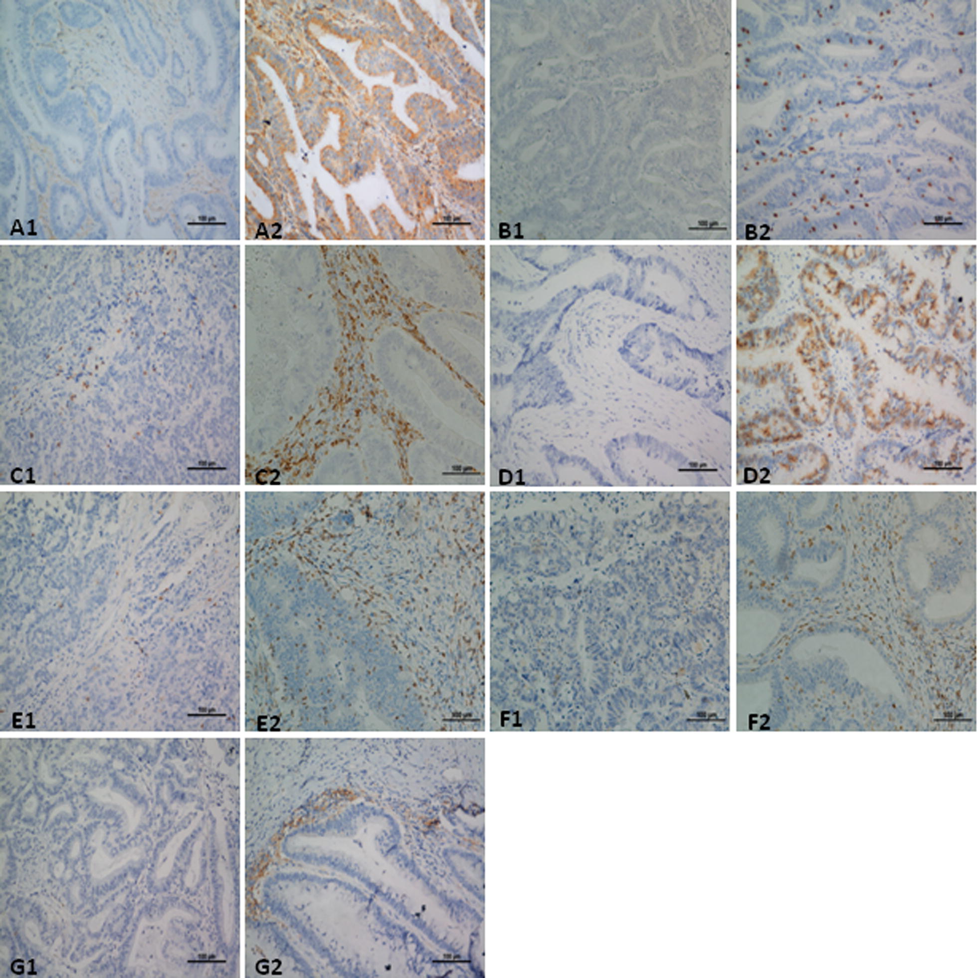
Fig. 1.

Representative IHC staining of immune molecules in CRC tissues. MHC class I expressed on tumor cells; CD3, CD4, CD8 and CD56 expressed in both intratumoral and interstitial areas; PD-1 expressed on lymphocytes; and PD-L1 expressed on tumor cells and tumor-infiltrating immune cells were evaluated. Low and high expression of MHC class I (A1 score 1, A2 score 3), CD3 (B1 score 1, B2 score 2), CD4 (C1 score 1, C2 score 3), CD8 (D1 score 0, D2 score 3), CD56 (E1 score 1, E2 score 3) and PD-1 (F1 score 0, F2 score 3); Negative and positive expression of PD-L1 (G1, G2). Images represent 200× microscopic fields used for analysis
