Fig. 1.
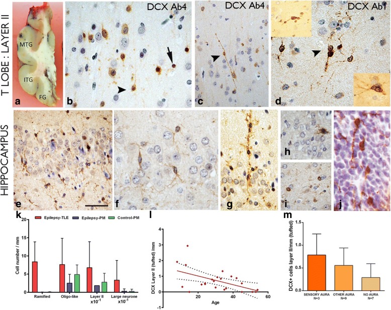
Doublecortin (DCX) in the cortex and hippocampus. a Section though a temporal lobe indicating the regions studies (MTG = middle temporal gyrus, ITG, inferior temporal gyrus, FG = fusiform gyrus) b Layer II DCX positive cells (DCX+) using DCX Ab 4 (see Table 2). Cells of different size, including some with more neuronal features and radial perpendicular processes (arrowhead) as well as dense nuclear labelling of small cells without processes (arrow) were observed. C. A bipolar cell in cortical layer II with DCX labelling with long beaded processes extending perpendicularly into layer I. d Clusters of small, intensely labelled DCX+ cells at interface of layer II and I labelled using DCX Ab1 (see Table 2). Top insert shows clusters of DCX+ cells; the bottom insert shows prominent nucleoli and neuronal appearance of DCX+ cells. e In the hippocampus granule cell layer (GCL) small DCX+ cells with ramified, multiple processes were observed; f In another case, the delicate branching processes of the ramified cells are shown. g A column of DCX+ cells extending though the GCL was observed in another case. h Granule cell neurons showed occasional DCX expression. i Small round DCX+ oligo-like cells were noted in the hippocampus in satellite location to neurons. j.DCX expression, in the periventricular germinal matrix of the lateral ventricle, in a developmental human control of 13 weeks, showing small cells with extended processes. k Bar chart showing greater linear densities for all morphological DCX+ cell types in surgical epilepsy cases compared to post mortem (PM) epilepsy controls and controls with statistically significant differences noted for ramified cell types only (p < 0.0001). l The linear density of layer II tufted DCX+ cell types showed an inverse correlation with age for all cases (surgical and PM) (p = 0.001) as well as for surgical cases alone (p = 0.016; not shown on graph). m Although greater DCX+ linear densities of tufted cells were present in patients with sensory aura of abnormal taste and smell compared to other aura types or no auras but these differences were not significant. Bar = 1 cm in A; B, D, F, G-J = 20 μm approx. (original magnification × 400) and C and E = 50 μm approx

