Fig. 3.
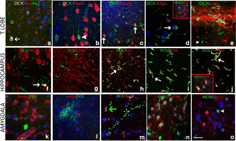
Characterizing DCX positive cells in the temporal cortex, hippocampus and amygdala of surgical patients with epilepsy, and comparison of commercial antibodies. In all panels, the arrowheads indicate single labelled cells, while arrows point to double labelled cells. Confocal images are merged projections of 5 to 7 images acquired in a z-stack. a. The immunoreactivity of two different commercially-available anti-Dcx antibodies. Both DCX Ab2 AB18723 (Abcam, Cambridgeshire, UK) and DCX Ab 1 #4606 (Cell Signaling Technology, Inc. MA, USA) labelled small cells in the hippocampal granule cell layer (GCL) of a patient with epilepsy and HS Type 1 (arrow) (Table 2). DCX Ab1 labelled more cells overall than DCX Ab2. DCX+ positive cells in the remaining figures were labelled using DCX Ab1. Temporal Cortex Layer I/II: b. NeuN expression was not frequently observed in small DCX+ cells located in layer II of the temporal lobe cortex (arrowhead). c. In another case, DCX/NeuN positive cells were more frequently observed in the superficial temporal cortex than in the temporal pole (arrows). d. DCX positive cells expressing Olig2 in the nucleus (arrow) were noted in layer 1 of an epilepsy case (arrow), but MCM2/DCX+ cells were not observed (inset). e. Nestin+ glial fibres were observed in the subpial layer and layer I and II, but did not co-localise with DCX expression. Hippocampus: f. In general there were rare DCX/NeuN colocalised cells in the dentate gyrus; in this image there is a rare co-localised cell (arrow). g. GFAP showed dense labelling of astroglial process in hippocampal regions but no co-localisation with DCX was noted. h. Labelling with Iba1 highlighted mature microglial cell types, with ramified processes, particularly in the subgranular zone as shown, and many co-expressed DCX (arrow); i Co-labelled CD68/DCX+ cells were also observed (arrow). j. PDGFRβ was expressed in multipolar cells in the hippocampus and temporal lobe in addition to pericytes; focal co-labelling with DCX was noted in some cells (arrow); Inset show a DCX+ ramified cell expressing MCM2 in the nucleus. Amygdala. k. Distinct populations or clusters of nestin+ or DCX+ cells in the amygdala periventricular nuclei; l. In another area, nests of DCX+ processes were also distinct from nestin-expressing cells. m. In the amygdala, although DCX+ processes were largely Iba1 negative, Iba1/DCX+ ramified DCX+ cells were observed (arrow). n. PDGFRβ and DCX in the amygdala showing distinct populations of small cells. o. DCX in the amygdala were mainly MCM2 negative. Bar is equivalent to 20 μm. Individual channels for immunofluorescence images are shown in Additional file 4: Figure S2

