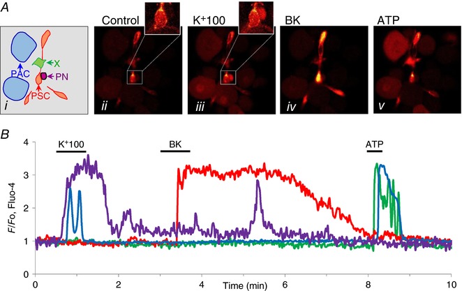
Figure 1. Simultaneous recordings of [Ca2+]i changes in response to various stimuli in four different cell types in a mouse pancreatic lobule.

Ai, sketch of location of different cell types in the lobule: blue, PACs; orange/red, PSCs; purple, PN; green, unknown (X). Aii–v, fluorescence images in control and during stimulation with high K+ (100 mm), BK (1 nm) and ATP (100 μm). As also seen in the [Ca2+]i traces shown in B, PN and PACs displayed rises in [Ca2+]i in response to membrane depolarization. PSCs responded to BK and both PACs and X responded to ATP. The colours of the traces in B match the coloured arrows in Ai.
