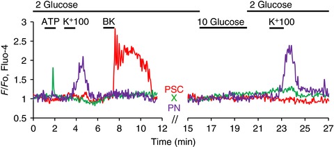
Figure 2. Elevating the extracellular glucose concentration from 2 to 10 mm has no effect on [Ca2+]i in any of the peri‐acinar cells.

In this experiment the lobule preparation was superfused with a solution containing 2 mm glucose, which then only late in the experimental protocol was replaced by 10 mm glucose for a few minutes. As shown in the green trace, an X‐cell responded to ATP (100 μm) with a rise in [Ca2+]i, but did not respond to subsequent challenges with high K+ (100 mm), BK (1 nm) or 10 mm glucose. In contrast, the PSC (red trace) did not respond to ATP or high K+, but only to BK. The PSC also failed to respond to the stimulation with 10 mm glucose. The PN (purple trace) only responded, repeatedly, to the high‐K+ stimulus.
