
Figure 1.
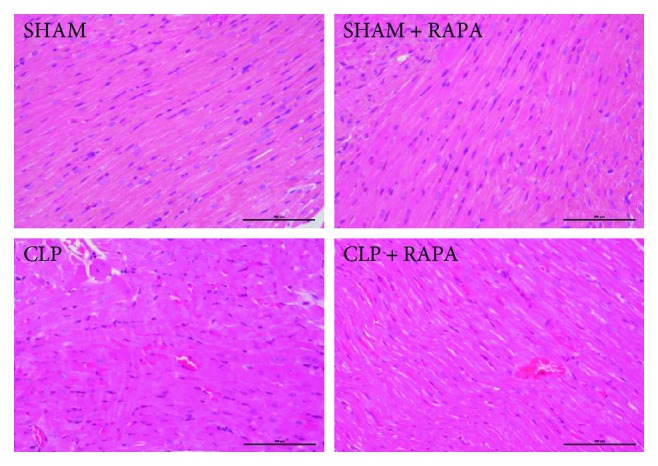
Representative H&E staining of the left ventricle sections. Original magnification, ×200. SHAM, sham operated; CLP, cecal ligation and puncture; RAPA, rapamycin.
Official websites use .gov
A
.gov website belongs to an official
government organization in the United States.
Secure .gov websites use HTTPS
A lock (
) or https:// means you've safely
connected to the .gov website. Share sensitive
information only on official, secure websites.
Representative H&E staining of the left ventricle sections. Original magnification, ×200. SHAM, sham operated; CLP, cecal ligation and puncture; RAPA, rapamycin.