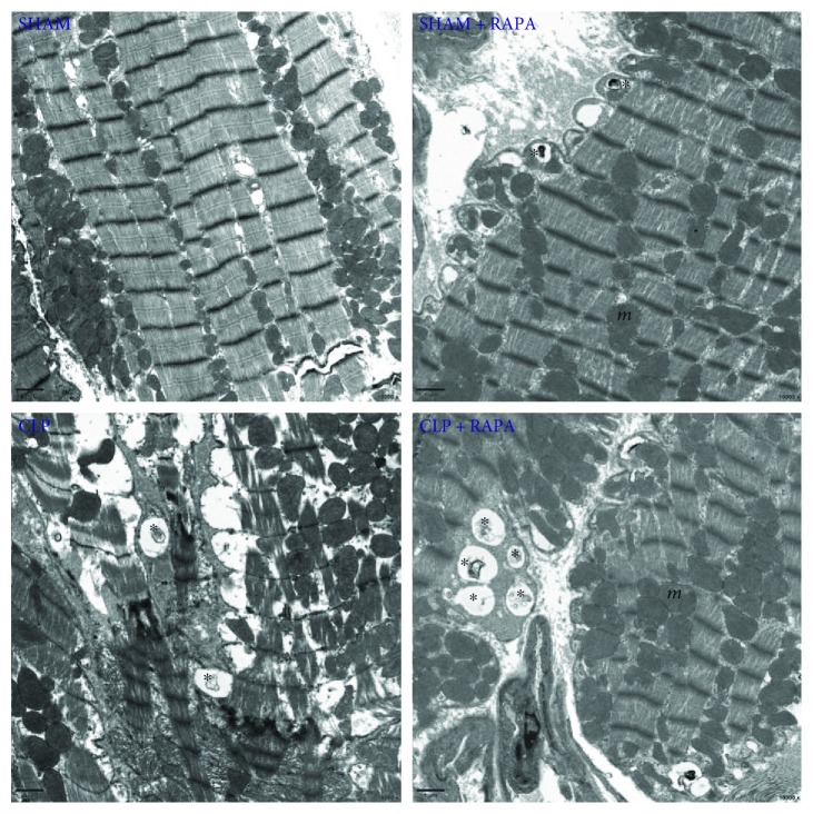
Figure 6.
Ultrastructural features of autophagic vacuoles in the left ventricle harvested 18 h after CLP. The myocardium was normal in appearance with a proper mitochondria distribution in sham-operated rats. Myofibrillar disarray was seen in CLP rats. There were more autophagic vacuoles (asterisk) in CLP + RAPA rats compared with CLP rats. Mitochondria (m) were seen throughout the cytoplasm. Magnification ×10,000.

