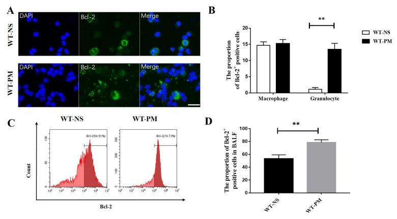
Figure 2.
PM increased the expression of Bcl-2 in BALF inflammatory cells. We counted the Bcl-2 positive cells in BALF cells by immunofluorescence assays between WT-NS mice and WT-PM mice (scale bar=20 μm). Cells with segmented nucleus were considered as granulocytes. Data are mean/SEM from 5 to 7 independent experiments, n>200 (A and B). BALF cells were isolated from WT-PM mice, and intracellular Bcl-2 expression was assessed by flow cytometry. The percentage of Bcl-2-positive cells is higher than WT-NS in BALF (C and D).

