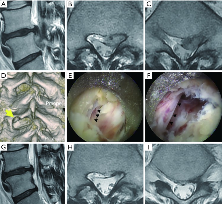
Figure 4.
Pre- and postoperative radiographic and intraoperative findings in a patient with highly down-migrated lumbar disc herniation (case 3). (A-C,G-I) Pre- (A-C) and postoperative (G-I) magnetic resonance imaging findings: sagittal (A,G) and axial (B,C,H,I) views of the T2-weighted image; (D) three-dimensional computed tomography: arrows indicate minimal laminectomy of the caudal margin of the left L5 vertebral laminae; (E,F) intraoperative photographs before (E) and after (F) the removal of the sequestrated nucleus. Arrowheads indicate caudal margin of the left L5 nerve root.

