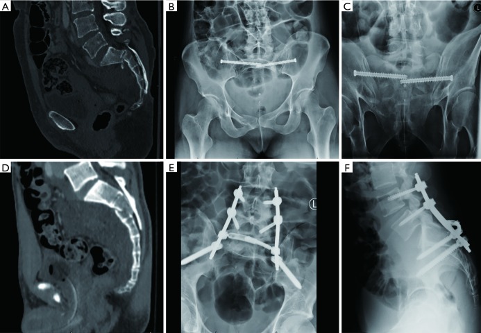
Figure 1.
(A) Sagittal CT cut and (B,C) post-operative inlet and outlet X-rays of a 51-year-old woman who underwent iliosacral screw fixation after sustaining a U-type sacral fracture after falling from a horse; (D) sagittal CT cut and (E,F) post-operative AP and lateral X-rays of a 26-year-old man who underwent lumbopelvic fixation after sustaining a U-type sacral fracture after sustaining a fall from a 30 ft roof. CT, computed tomography.

