
Figure 1.
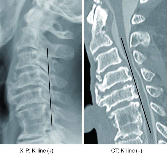
Representative cervical radiographs and CT-MPR mid-sagittal images of a same patient with cervical ossification of the longitudinal ligament. The X-P based K-line was found to be (+) (A), whereas the CT-based K-line was found to be (−) (B). The K-line was different between radiographs and the CT-MPR. CT-MPR, computed tomography-multiplanar reconstruction.