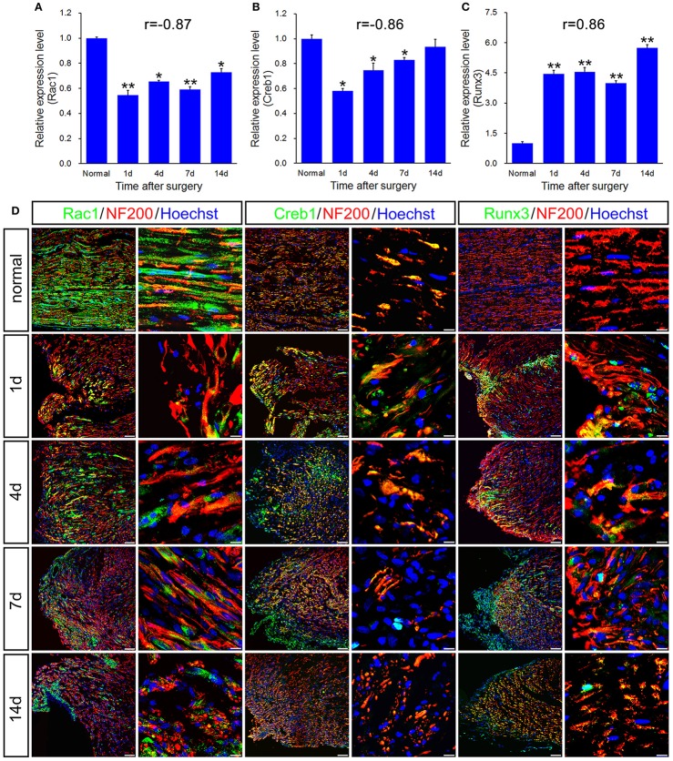
Figure 5.
qPCR and histological validation of selected transcription regulators and enzyme for both immune response and axonal regeneration following sciatic nerve transection. (A–C) Histograms showing the qPCR validation for relative mRNA expressions of Rac1, Creb1 and Runx3. The relative level was normalized to GAPDH. The data, obtained from 3 independent experiments, are expressed as means ± SEM. One-way ANOVA and post-hoc Bonferroni t-test were used to analyze the data. *P < 0.05, **P < 0.01 vs. the normal control. (D) Immunostaining by anti-Rac1, anti-Creb1 and anti-Runx3 (green) merged with anti-NF-200 (red) primary antibodies and Hoechst 33342 (blue) obtained at 1, 4, 7, 14 days post-sciatic nerve transection and normal sample of the longitudinal sectioned proximal nerve stump, respectively. Hoechst 33342 marks for nucleus and NF200 markers for nerve fibers. “r” stands for the correlation coefficient. Scale bar, 75 μm (low magnification) and 10 μm (high magnification).

