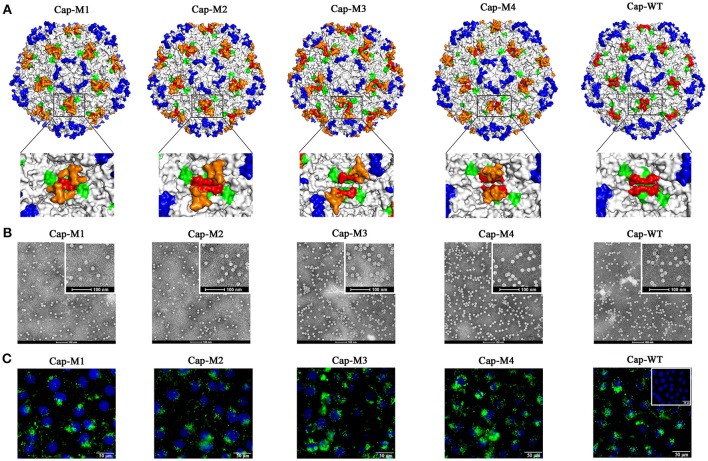
Figure 3.
PCV2 VLPs assembly and cell entry into PK15 cells. (A) Localizations of foreign peptides or epitopes on the surface of PCV2 capsid in simulated 3D models after assembly of PCV2 Cap-WT and mutants into VLPs. Images on bottom of (A) demonstrate (high magnification) 3D structures or orientations of foreign epitopes and artificial peptides on a plateau formed by two neighboring MP-Lcd. Foreign epitopes or artificial peptides were labeled in orange; MP-Lcd was labeled in red and Loop CD, except MP-Lcd was labeled in green and Loop BC-decorated five-fold axes of the icosahedra were labeled in blue on PCV2 capsid. (B) PCV2 VLPs formations observed by TEM. Negative staining of VLPs assembled from PCV2 Cap WT or mutants (bars = 100 nm). (C) Entry of PCV2 VLPs into PK-15 cells. Internalizations of PCV2 VLPs assembled from PCV2 Cap WT or Cap mutants (M1-M4) were confirmed by confocal microscopy. Inset in PCV2 Cap-WT represents a negative control in which PBS instead of VLPs was added into PK15 cell culture. Green fluorescence represents PCV2 Cap in PK15 cells. Nuclei (blue) of PK-15 cells were stained by DAPI.

