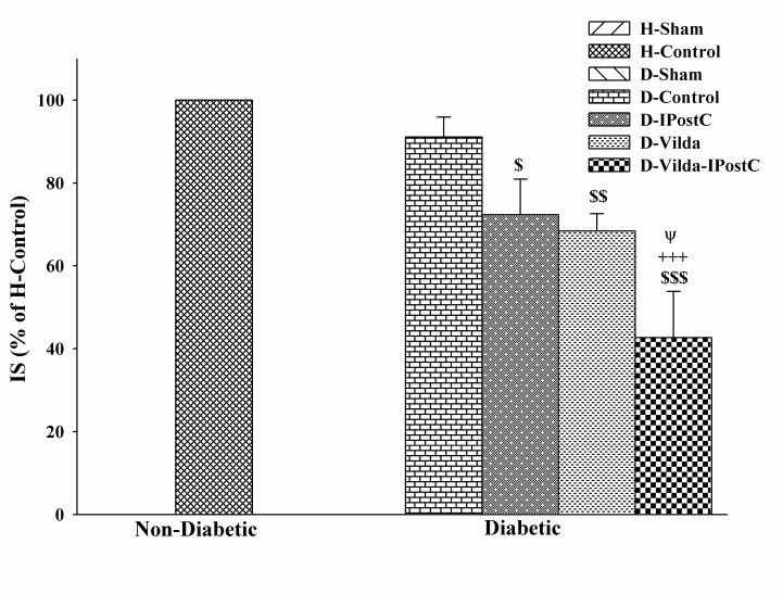
Figure 1.
The changes of infarct sizes (IS) in experimental groups. IS was evaluated by evan's blue and triphenyltetrazolium chloride staining method, in non-diabetic and diabetic hearts subjected to 35 min regional ischemia and 60 min reperfusion. The data were expressed as mean±SEM. n=5-6 for each group. H: healthy; D: diabetic; IPostC: ischemic postconditioning; Vilda: vildagliptin. $p<0.05, $p<0.01 and $p<0.001 as compared to H-control;+++p<0.001 as compared to D-Control group; Ψp<0.05 as compared to D-Vilda and D-IPostC groups.

