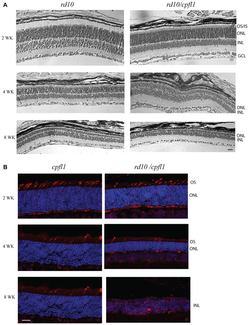
Figure 1.
Histological analysis of rd10/cpf1 mouse retinas. (A) Comparison of morphology of rd10/cpfl1 to rd10 by H&E staining. Rod photoreceptor cells degenerate with age as shown by thinning of ONL and loss of photoreceptor outer and inner segments. (B) Comparison of cone photoreceptor cell degeneration in rd10/cpfl1 with cpfl1 by anti-cone opsin antibody staining (labeled as red). OS/IS, outer/inner segments; ONL, outer nuclear layer; INL, inner nuclear layer; GCL, ganglion cell layer. Scale bar: 20 μm.

