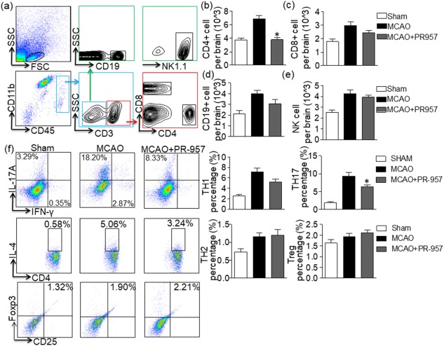
Figure 2.

PR‐957 decreases the number of CD4+ T cells in the brain after middle cerebral artery occlusion (MCAO) and suppresses T helper type 17 (Th17) differentiation. At 72 h after MCAO, cells were harvested from the brains of mice treated with PR‐957 (MCAO + PR‐957), vehicle (MCAO + vehicle) and sham operation (sham), and flow cytometric analysis was conducted. (a) Gating strategy for leucocytes isolated from the brain. (b–e) Counts of CD4+ T cells (CD3+CD4+), CD8+ T cells (CD3+CD8+), natural killer (NK) cells (CD3−NK1.1+), and B cells (CD3−CD19+) in the indicated groups. Flow cytometric analysis showed a significant decline in CD4+ T cells in the mice treated with PR‐957 group compared to the group treated with vehicle. PR‐957 had no significant effect on CD8+ T, B or NK cells. Dot‐plots representing the distribution of different cell types counted during fluorescence activated cell sorter (FACS) for individual cytokines. The areas enclosed in boxes indicate the percentages of CD4+interferon (IFN)‐γ+ cells, CD4+interleukin (IL)‐17+ cells, CD4+IL‐4+ cells and forkhead box protein 3 (FoxP3+) CD25+ cells among the CD4+ T cells, which are presented quantitatively in the bar graphs of panel (f). Data are presented as means ± standard error of the mean (s.e.m.); # P < 0·05, compared with sham; *P < 0·05, compared with MCAO + vehicle; n = 6 mice per group. The experiment was repeated twice with similar results.
