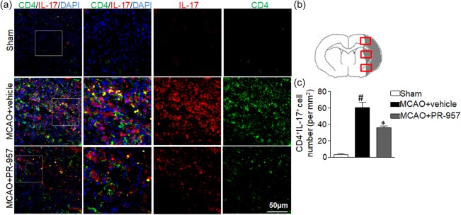
Figure 3.

PR‐957 lessens T helper type 17 (Th17) cell infiltration in brains of middle cerebral artery occlusion (MCAO) mice. (a) Brain sections from the three groups of mice were stained for interleukin (IL)‐17 and CD4 using double‐fluorescent immunohistochemistry to analyse Th17 cell brain infiltration after MCAO [4′,6‐diamidino‐2‐phenylindole (DAPI) counterstain]. (b) Schematic representation of a coronal brain section with analysis fields overlain in the cortex and striatum, from which Th17‐positive cells per mm2 were counted. In the sham‐operated group, the IL‐17‐positive staining was difficult to detect as very few IL‐17‐positive cells had infiltrated. However, in the ipsilateral ischaemic hemisphere 72 h after MCAO, IL‐17 cells became activated. The PR‐957‐treated group had fewer Th17‐positive cells compared with the MCAO + vehicle group. (c) Quantitation of brain‐infiltrated Th17 cells in the indicated groups. There were significantly fewer Th17‐positive cells in the PR‐957‐treated group than in the MCAO + vehicle group. Bars and error bars represent means ± standard error of the mean (s.e.m.); # P < 0·05, compared with sham‐operated; *P < 0·05, compared with MCAO + vehicle; n = 6 mice in each group. Experiments were repeated three times, yielding similar results. Same labelling conventions as previous figures.
