Figure 1.

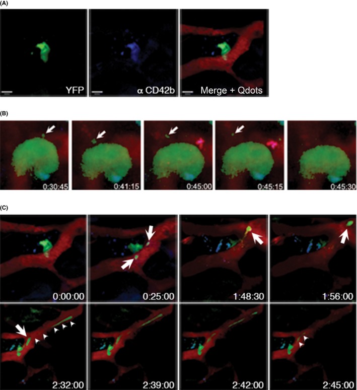
Platelet biogenesis in vivo. (A) eYFP+‐MKs are detected in MPM (left panel) and are additionally labeled by anti‐CD42b‐antibodies (middle panel). Vessels and sinusoids are visualized by intravenous injection of Qdots‐655 (right panel). Scale bars indicate 50 μm. (B) Still images from time‐lapse microscopy video 1 hour after antibody injection. Antibody decorated MKs are able to form proplatelets (arrows). (C) Still images from a video showing that antibody‐decorated MKs are able to form platelet‐sized particles as well as long, extended protrusions (proplatelets, arrows), which grow substantially within the overall observation period (arrow‐heads, lower panel)
