
Fig. 3.
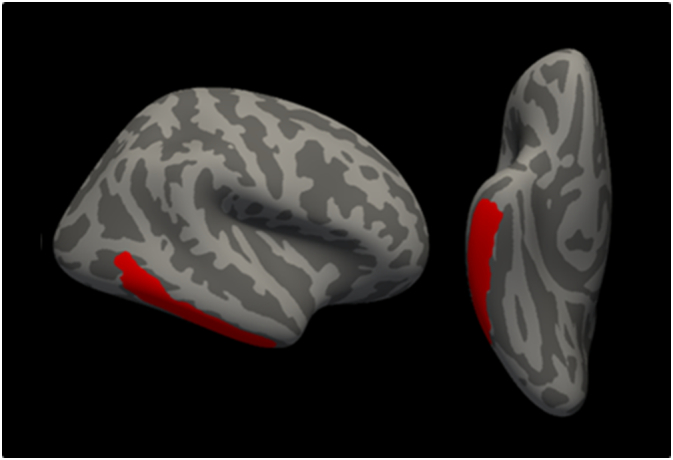
Distribution of the cortical thinning (in red) on the pial surface of the right hemisphere (lateral and inferior views) in ALSimp patients compared to ALScn patients. (For interpretation of the references to colour in this figure legend, the reader is referred to the web version of this article.)