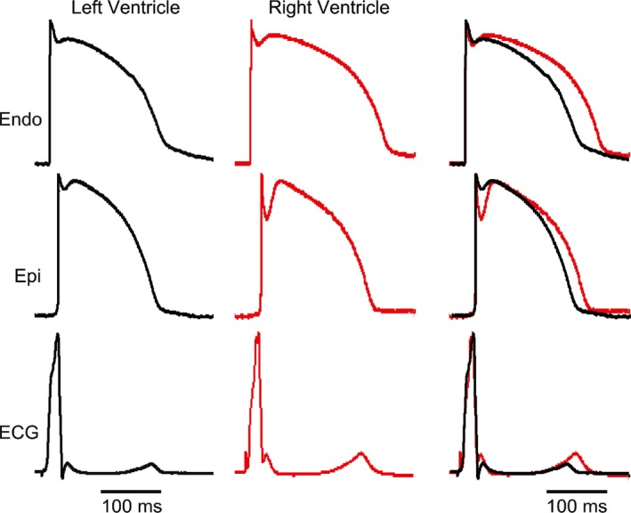
Figure 1.

Representative recordings of endocardial (endo) and epicardial (epi) action potentials (APs) and the corresponding transmural‐ECG (ECG) obtained from a RV and LV preparation. Preparations were stimulated from the endo surface at a BCL = 1 sec. Although LV preparations were thicker than RV preparations, the superimposed ECGs show that transmural conduction time was similar.
