Figure 3.

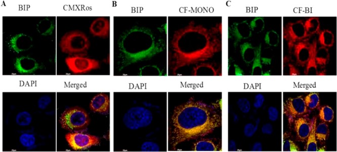
Mitochondrial staining by Bodipy fluorophores in HeLa cells: The cells were incubated with Bodipy fluorophores (100 nM) at 37 °C for 30 min. (A) Green fluorescence indicates mitochondrial staining of BIP compound by confocal microscopy using FITC filter. Red fluorescence indicates mitochondrial staining of Mitotracker Red CMXRos (Thermofisher) by confocal microscopy by using the rhodamine filter. Blue fluorescence indicates nuclear staining by DAPI. (B) Same as A, except that red fluorescence indicates mitochondrial staining of CF-MONO compound by confocal microscopy by the using rhodamine filter. (C) Same as A, except that red fluorescence indicates mitochondrial staining of CF-BI compound by confocal microscopy by using the rhodamine filter.
