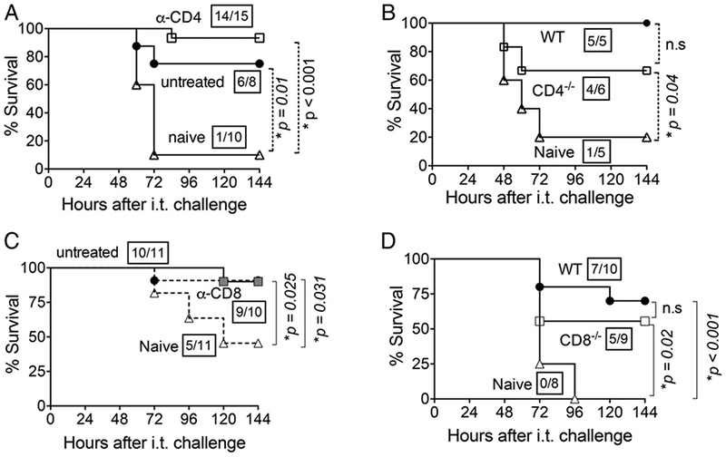
FIGURE 6.

CD4+ and CD8+ T cells are individually dispensable for NP exposure-mediated protection. (A) Three weeks after NP exposure of 8- to 10-wk-old C57BL/6 mice, they were treated with 0.5 μg of GK1.5 anti-CD4 mAb at days −2, −1, and +1 relative to i.t. challenge to deplete 98% of CD4+ T cells (data not shown). Survival of CD4-depleted mice (α-CD4; □), untreated NP-exposed C57BL/6 mice (●), and naive C57BL/6 controls (Δ) was assessed serially after i.t. challenge with 2 × 10⁶ CFU of S. pneumoniae TIGR4. (B) Survival of NP-exposed CD4-deficient BALB/c mice (□), NP-exposed wild-type (WT; ●), or naive wild-type mice (naive; Δ) was assessed after i.t. challenge with 2 × 10⁶ CFU of S. pneumoniae TIGR4. (C) Three weeks after NP exposure, wild-type BALB/c mice were treated with 0.5 μg of clone 2.43, an mAb against CD8, at days −2, −1, and +1 relative to i.t. challenge to deplete 98% of CD8+ T cells (data not shown). Survival of CD8-depleted mice (α-CD8; ■) was assessed at indicated time points after i.t. challenge with 2 × 10⁵ CFU of S. pneumoniae TIGR4 and compared with NP-exposed, nondepleted mice (●) and naive controls (Δ). (D) Three weeks after NP exposure, CD8-deficient mice in BALB/c background (CD8–/–; □) and wild-type BALB/c mice (WT; ●) or naive BALB/c wild-type mice (naive; Δ) were i.t. challenged with 1 × 10⁶ CFU of S. pneumoniae TIGR4. Survival was assessed at the indicated time points after challenge and compared with naive controls (□). For (A)–(D), fractions denote survivors 2 wk after challenge over the total number of mice, and asterisks indicate statistical significance (p < 0.05) by the log-rank (Mantel–Cox) test. Data pooled from two independent experiments are shown. n.s., not significant.
