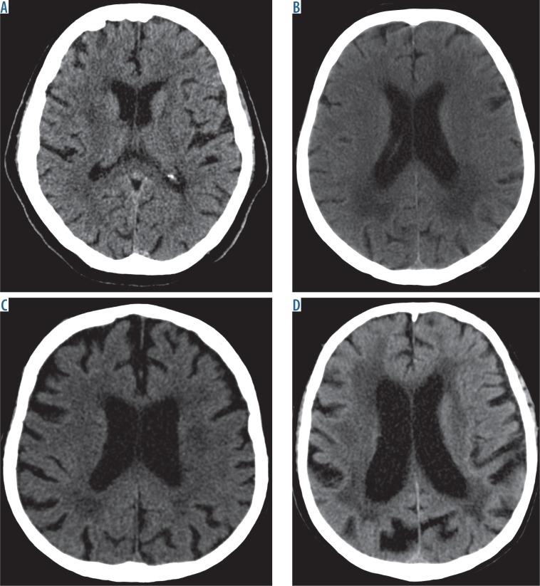
Fig. 1.
Computed tomography brain images at the level of the ventricular system show the respective grades of severity of leukoaraiosis. A) Grade 1 on the scale by van Swieten – small hypodense lesions can be seen around the anterior horns of the lateral cerebral ventricles. B) Grade 2 on the scale by van Swieten – diffuse, confluent hypodense lesions can be seen around the posterior horn of the lateral cerebral ventricles that extend to the cerebral cortex. C) Grade 3 on the scale by van Swieten – lesions extending to the subcortical areas are seen in the posterior horns of the lateral ventricles; leukoaraiotic lesions are less pronounced in the anterior part of the brain. D) Grade 4 on the scale by van Swieten – diffuse hypodense lesions are seen around the ventricles and in the semioval centres

