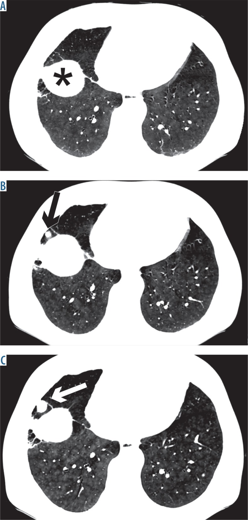
Figure 3.
Follow-up computed tomography (CT) examinations. Slight progression of diffuse, centrilobular ground-glass opacities is visible bilaterally in the lower parts of the lungs due to metastatic pulmonary calcification, assessed retrospectively – (A) baseline CT, (B) follow-up CT after 1 year, (C) follow-up high-resolution chest computed tomography after 2 years (arrows show progression of metastases of renal carcinoma, *diaphragm)

