Fig. 6.
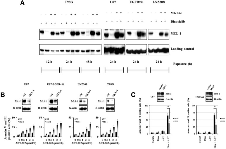
Dinaciclib promotes proteasomal degradation of Mcl-1 and enhances ABT-737-mediated cell death in malignant human glioma cell lines. (A) Logarithmically growing T98G, U87, U87-EGFRviii, and LNZ308 cells were pretreated with 1.0 µM of MG-132 (proteasomal inhibitor) for 2 hours followed by dinaciclib (250 nM) for the indicated duration. Cell extracts were subjected to Western blot analysis with the indicated antibody. β-actin served as loading control. (B) U87, U87-EGFRviii, LNZ308, and T98G cells were transfected with nontarget (NT) or Mcl-1 shRNA as described in Materials and Methods. Forty-eight hours post-transfection, cells were treated with the indicated concentrations of ABT-737 for 24 hours, and viability was assessed by annexin V/PI apoptosis assay (lower panel). In parallel, cell lysates were collected and protein was subjected to Western blot analysis using Mcl-1 antibody. Immunoblots were stripped and reprobed with β-actin. (C) U87 and LNZ308 cells were transfected with vector (pCMV) or Mcl-1 expression vector as described in Materials and Methods. Forty-eight hours post-transfection, cells were treated with dinaciclib (dina, 100 nM) or ABT-737 (ABT, 100 nM) or the combination of both (dina + ABT) for 24 hours, and viability was assessed by annexin V/PI apoptosis assay (lower panel). In parallel, cell lysates were collected, and protein was subjected to Western blot analysis using Mcl-1 antibody. Immunoblots were stripped and reprobed with β-actin. Data are representative of triplicate studies from three independent experiments. *P < 0.005.

