
Extended Data Figure 5. Microglia activation in the brain starts at early, preclinical stages.
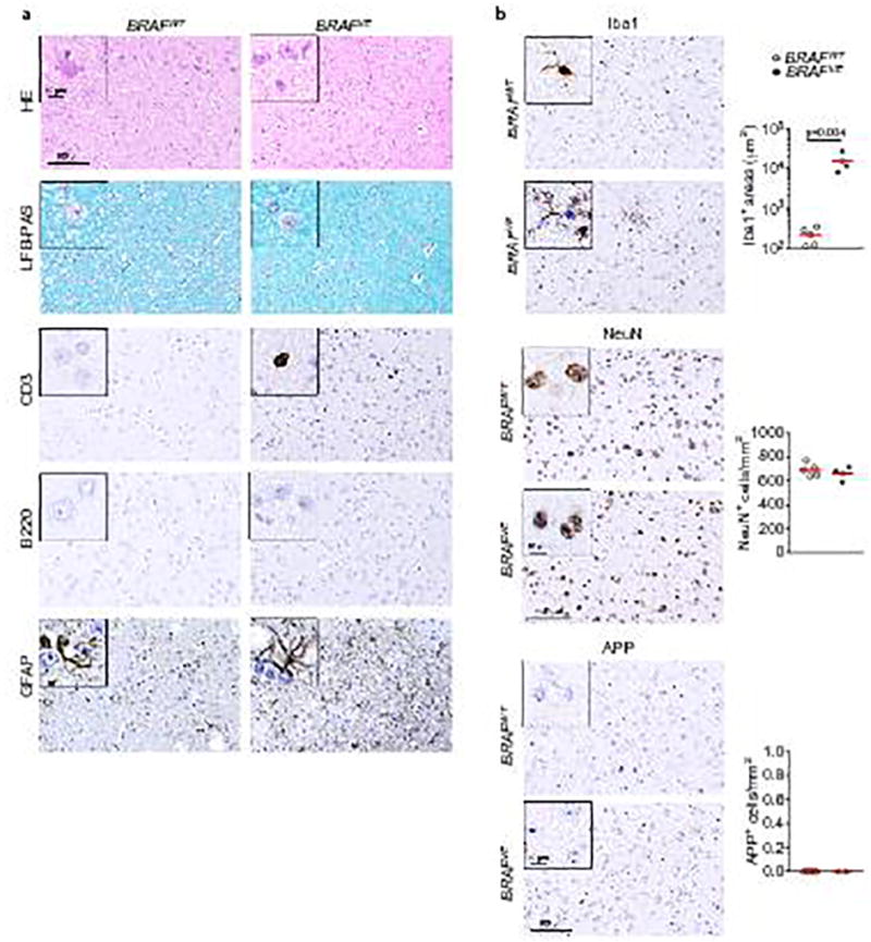
(a) Histological analysis by hematoxylin and eosin (HE) and Luxol-fast-blue-PAS (LFB-PAS) and immunohistochemistry analysis of T-cells (CD3), B-cells (B220) and astrocyte activation (GFAP) in one-month old BRAFVE mice and BRAFWT littermates. Representative of n=5 per for BRAFWT and n=4 for BRAFVE. (b) Immunohistochemistry analysis and quantification of Iba1+ cell density, cortical neurons (NeuN) and expression of amyloid precursor protein (APP), a positive signal for neurodegeneration in one-month old BRAFVE mice and BRAFWT. Representative of n=5 per for BRAFWT and n=4 for BRAFVE. Circles represent individual mice. Scale bars=100µm (10µm for insets). Unpaired two-tailed t-test.