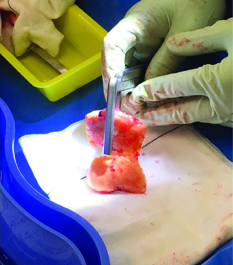
Figure 3.
Measurement of the medial and lateral distal femoral condyle and posterior condylar cuts. Sequential pinning of the block was carried out very carefully with 2 distal pins (through the 3D block) followed by 2 anterior femoral pins (through the in situ jig). The oblique anterior femoral pin was placed last to prevent any movement of the 3D block whilst resecting. Distal femoral resections were then done through the in situ jig. The posterior condylar cuts were done through the appropriate standard 4:1 in block as per plan. The largest thickness of bony prominence was recorded by the senior surgeon using Vernier Calipers for each resected medial and lateral distal femoral condyle. Three separate measurements − horizontal, vertical and diagonal were made, and the average measurement was recorded independently.

