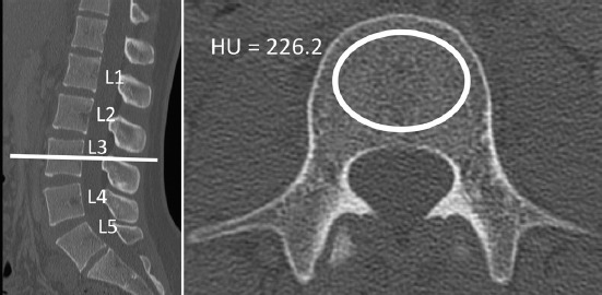
Figure 2.

CT attenuation was measured by first locating the mid-vertebral body in the sagittal plane. Axial click-and-drag elliptical ROI were manually placed to be as large as possible while safely avoiding the cortical shell and vertebrobasilar complex. The picture archiving software reported the average CT attenuation of the ROI in Hounsfield units (HU)
