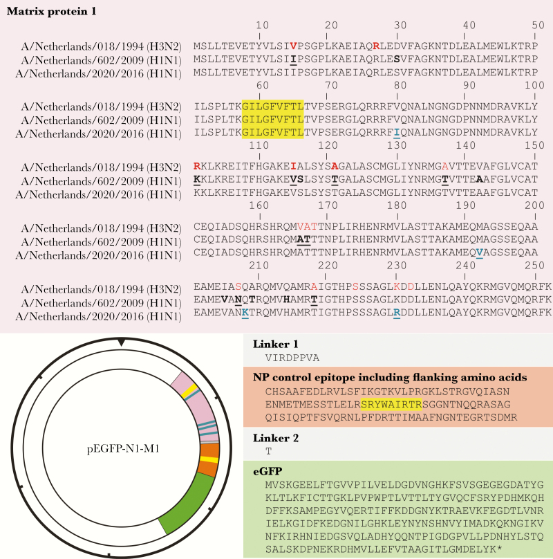
Figure 1.
Amino acid sequences of viral matrix protein 1-nucleoprotein-enhanced green fluorescent protein (M1-NP-eGFP) fusion proteins and expression plasmid map. The amino acid sequence of the chimeric M1-NP-eGFP fusion construct is shown: M1 encoding sequences A/Netherlands/018/1994 (H3N2s1994), A/Netherlands/602/2009 (H1N1pdm09), and A/Netherlands/2020/2016 (H1N1pdm09/2016) (pink), NP (orange), eGFP (green), and linker sequences (gray), and M158-66 (GILGFVFTL) and NP383-391 (SRYWAIRTR) epitopes (yellow). Previously identified extra-epitopic amino acid residues of a human signature are indicated in bold red and additional extra-epitopic amino acid residues of a human signature are indicated in red [10]. Amino acid difference between the H3N2s1994 and H1N1pdm09 M1 proteins are indicated in bold, and those of avian/swine signature are bold underlined. Amino acid differences studied in this report between H1N1pdm09 and H1N1pdm09/2016 M1 proteins are indicated in blue, bold and underlined. These inserts were cloned into the pEGFP-N1 vector as indicated. The hash marks around the perimeter of the plasmid map indicate 1000-nucleotide increments.

